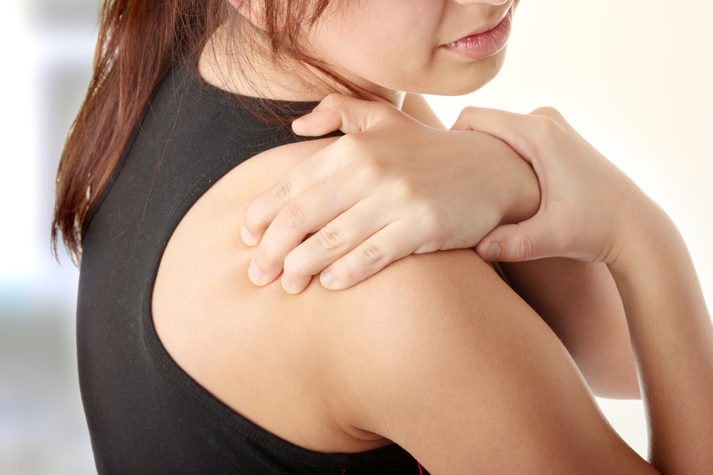
Shoulder impingement syndrome, also called painful arc syndrome, , swimmer’s shoulder, and thrower’s shoulder, is a clinical syndrome which occurs when the tendons of the rotator cuff muscles become irritated and inflamed as they pass through the subacromial space, the passage beneath the acromion . This can result in pain, weakness and loss of movement at the shoulder.
Causes
 When the arm is raised, the subacromial space (gap between the anterior edge of the acromion and the head of the humerus) narrows, through which the supraspinatus muscle tendon passes.[3] Anything that causes further narrowing has the tendency to impinge the tendon and cause an inflammatory response, resulting in impingement syndrome. This can be caused by bony structures such as subacromial spurs (bony projections from the acromion), osteoarthritic spurs on the acromioclavicular joint, and variations in the shape of the acromion. Thickening or calcification of the coracoacromial ligament can also cause impingement. Loss of function of the rotator cuff muscles, due to injury or loss of strength, may cause the humerus to move superiorly, resulting in impingement. Inflammation and subsequent thickening of the subacromial bursa may also cause impingement
Sport-Specific Biomechanics
- Overuse or repetitive microtrauma sustained in the overhead position may contribute to impingement and rotator cuff pathology. Shoulder pain and rotator cuff disease are common in athletes involved in sports requiring repetitive overhead arm motion (eg, swimming, baseball, volleyball, tennis).
- Secondary impingement often is attributed to impingement, which seldom is mechanical in nature in young athletes. Rotator cuff disease in this population may be related to subtle instability, and, therefore, may be secondary to such factors as eccentric overload, muscle imbalance, glenohumeral instability, or labral lesions. This has led to the concept of secondary impingement, which is defined as rotator cuff impingement that occurs secondary to a functional decrease in the supraspinatus outlet space due to underlying instability of the glenohumeral joint.
- Secondary impingement may be the most common cause in young athletes who frequently place large, repetitive overhead stresses on the static and dynamic glenohumeral stabilizers, resulting in microtrauma and attenuation of the glenohumeral ligamentous structures, which leads to subclinical glenohumeral instability. Such instability places increased stress on the dynamic stabilizers of the glenohumeral joint, including the rotator cuff tendons.
- These increased demands may lead to rotator cuff pathology (eg, partial tearing, tendonitis). Furthermore, as the rotator cuff muscles fatigue, the humeral head translates anteriorly and superiorly, impinging upon the coracoacromial arch. This leads to rotator cuff inflammation. In these patients, treatment should address underlying instability.
Occurence
Age:
- Patients younger than 40 years – Usually glenohumeral instability, and acromioclavicular joint disease/injury
- Patients older than 40 years – Consider glenohumeral impingement syndrome/rotator cuff disease and glenohumeral joint degenerative disease
Occupation:
- Individuals at highest risk for shoulder impingement are laborers and those working in jobs that require repetitive overhead activity.
- Athletes (eg, swimming, throwing sports, tennis, volleyball)
Athletic activity:
- Onset of symptoms in relation to specific phases of the athletic event performed
- Duration and frequency of play
- Duration and frequency of practice
- Level of play (eg, little league, high school, college, professional)
- Actual playing time (eg, starter, backup, bench player) and position played
- Lack of periodization in training – Athlete participating in same overhead sport year-round
Symptoms
- Onset: Sudden onset of sharp pain in the shoulder with tearing sensation is suggestive of a rotator cuff tear. Gradual increase in shoulder pain with overhead activities is suggestive of an impingement problem.
- Chronicity of symptoms
- Location: Pain usually is reported over the lateral, superior, anterior shoulder; occasionally refers to the deltoid region. Posterior shoulder capsule pain usually is consistent with anterior instability, causing posterior tightness.
- Setting during which symptoms arise (eg, pain during sleep, in various sleeping positions, at night, with activity, types of activities, while resting)
- Quality of pain (eg, sharp, dull, radiating, throbbing, burning, constant, intermittent, occasional)
- Quantity of pain (on a scale of 0-10, 10 being the worst)
- Alleviating factors (eg, change of position, medication, rest)
- Aggravating factors (eg, change of position, medication, increase in practice, increase in play, change in athletic gear/foot wear, change in position played)
- Functional symptoms – Patient changed mechanics (eg, throwing motion, swim stroke) to compensate for pain
- Associated manifestations (eg, possibly chest pain, dizziness, abdominal pain, shortness of breath)
- Provocative position: Pain with humerus in forward-flexed and internally rotated position suggests rotator cuff impingement. Pain with humerus in abducted and externally rotated position suggests anterior glenohumeral instability and laxity.
Other history
Inquire about previous or recent trauma, stiffness, numbness, paresthesias, clicking, weakness, crepitus of instability, and neck syndromes.
Following are the 3 stages in the spectrum of rotator cuff impingement:
- Stage 1, commonly affecting patients younger than 25 years, is depicted by acute inflammation, edema, and hemorrhage in the rotator cuff. This stage usually is reversible with nonoperative treatment.
- Stage 2 usually affects patients aged 25-40 years, resulting as a continuum of stage 1. The rotator cuff tendon progresses to fibrosis and tendonitis, which commonly does not respond to conservative treatment and requires operative intervention.
- Stage 3 commonly affects patients older than 40 years. As this condition progresses, it may lead to mechanical disruption of the rotator cuff tendon and to changes in the coracoacromial arch with osteophytosis along the anterior acromion. Surgical l anterior acromioplasty and rotator cuff repair is commonly required
Treatment:
Acute Phase
Physical Therapy
Goals of the acute phase are to relieve pain and inflammation, prevent muscle atrophy without exacerbation, reestablish nonpainful ROM, and normalize arthrokinematics of the shoulder complex. A period of active rest should be recommended to the patient, eliminating any activity that may cause an increase in symptoms. ROM exercises may include pendulum exercises and symptom-limited active-assistive range of motion (AAROM) exercises. Joint mobilization may be included with inferior, anterior, or posterior glides in the scapular plane. Strengthening exercises should be isometric in nature, working on the external rotators, internal rotators, biceps, deltoids, and scapular stabilizers (rhomboids, trapezius, serratus anterior, latissimus dorsi, and pectoralis major).
Exercises targeting the rotator cuff muscles are extremely important. Neuromuscular control exercises also may be initiated. Modalities may be used as an adjunct and can include cryotherapy, transcutaneous electrical nerve stimulation (TENS), high-voltage galvanic stimulation, ultrasound, phonophoresis, or iontophoresis. Patient education is particularly important for the acute phase regarding activity, pathology, and avoiding overhead activity, reaching, and lifting. The general guidelines to progress from this phase are decreased pain or symptoms, increased ROM, painful arc in abduction only, and improved muscular function.
Subacromial injection
During the acute to subacute phase, when pain and inflammation are predominant, a subacromial injection may be diagnostic and therapeutic as an adjunct to a rehabilitation program. Injection of 10 mL of 1% lidocaine solution (without epinephrine) into the subacromial space should relieve shoulder pain if pain and inflammation truly is originating from the supraspinatus outlet/subacromial space. Adding a low dose intermediate-acting injectable corticosteroid may provide a therapeutic effect. Betamethasone, triamcinolone, and methylprednisolone commonly are used
High-intensity laser therapy
After 2 weeks, patients in the HILT group showed statistically significant improvements in pain reduction, articular movement, functionality, and muscle strength as measured by 3 outcome measure scores. However, further investigation is warranted, as the study was limited by its small size, lack of control or placebo groups, and follow-up period.
Surgical Intervention
In general, conservative measures are continued for at least 3-6 months or longer if the patient is improving, which is usually the case in 60-90% of patients. If the patient remains significantly disabled and has no improvement after 3 months of conservative treatment, the clinician must seek further diagnostic work-up, and reconsider other etiologies or refer for surgical evaluation.
Appropriate surgical referrals are patients with subacromial impingement syndrome refractory to 3-6 months of appropriate conservative treatment
Surgical evaluation
Initial examination under anesthesia (general anesthesia vs. regional block) and diagnostic arthroscopy
Evaluation of shoulder ROM and stability
In patients with limited motion, manipulation of the shoulder is performed. Diagnostic arthroscopy also may be performed, but arthroscopic subacromial decompression is generally not performed in patients with significant preoperative stiffness due to the increased risk of postoperative adhesive capsulitis.
Arthroscopic evaluation
Particular attention is directed to the rotator cuff, especially the supraspinatus tendon near its insertion onto the greater tuberosity
Medication
During the acute and subacute phases of shoulder impingement syndrome, it is appropriate to use a short course of nonsteroidal anti-inflammatory drugs (NSAIDs) for analgesic and anti-inflammatory effects as an adjunct to the therapy program and other treatment modalities
Return to Play
Return to play is restricted until full pain-free ROM is restored, both rest and activity-related pain are eliminated, and provocative impingement signs are negative. Isokinetic strength testing must be 90% compared to the contralateral side. When the patient is symptom-free, resuming activities is gradual, first during practice to build up endurance while working on modified techniques/mechanics, and then in simulated game situations. The athlete should continue flexibility and strengthening exercises after returning to his/her sport to prevent recurrence.
